Research-based patient information · age scope stated below
Brain and spinal tumours: finding the right care pathway
A tumour is an abnormal tissue growth; it is not automatically malignant. Location, behaviour, molecular diagnosis, pressure on the brain or spinal cord, overall health and the person's priorities all shape the plan. This overview links primary brain tumours, brain metastases and spinal-canal disease without treating them as one condition.
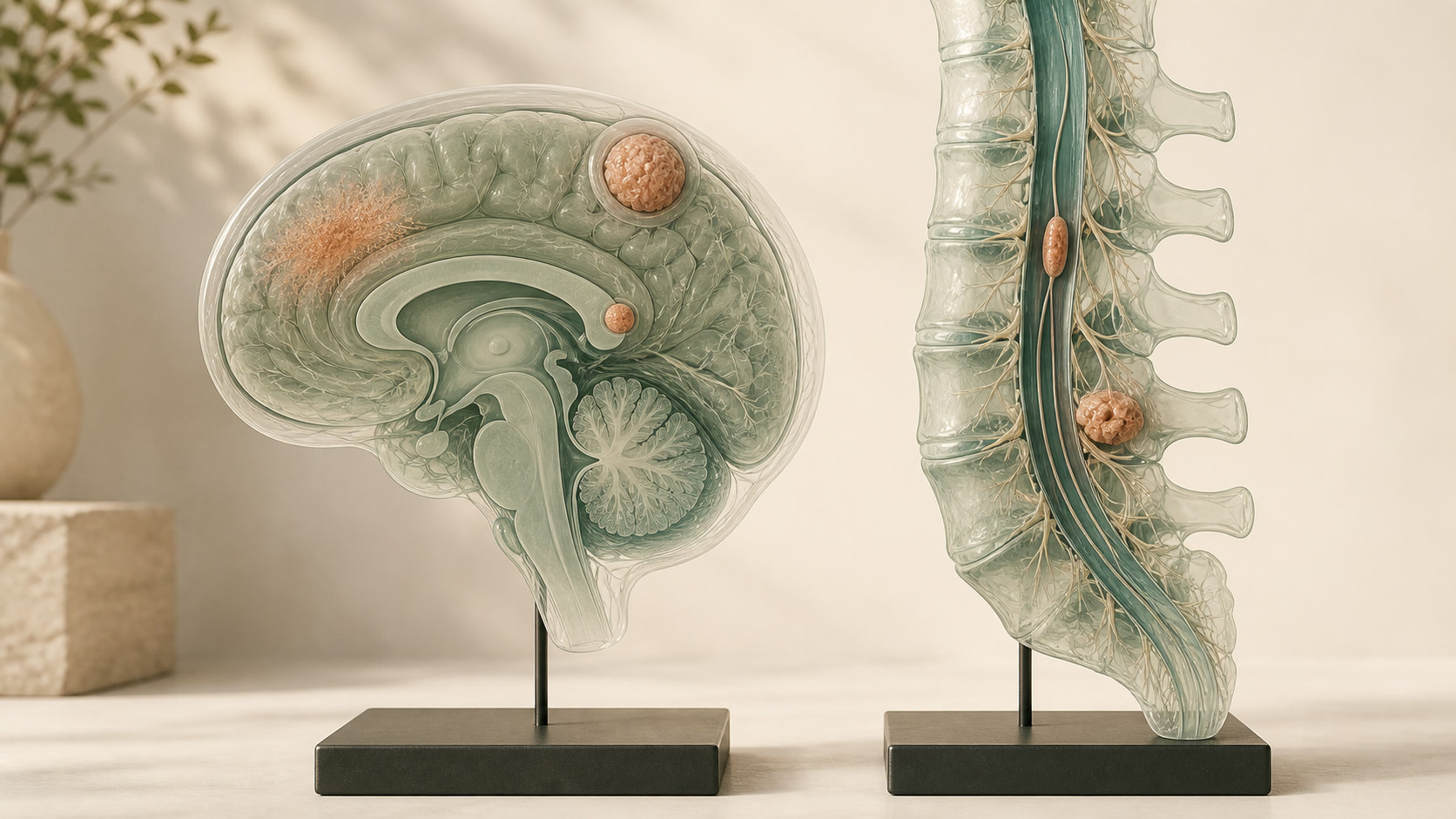
01
Different conditions need different pathways
- Glioma is a family of primary central-nervous-system tumours classified by an integrated assessment of tissue appearance and molecular features.
- Meningioma usually arises from the coverings of the brain or spinal cord; many are slow-growing and discovered incidentally.
- A brain metastasis has spread from cancer elsewhere in the body and is planned together with the wider cancer pathway.
- Primary spinal-canal or vertebral tumours and adult spinal metastases with possible metastatic spinal cord compression (MSCC) are distinct groups.
02
Symptoms depend on location
Possible features include a first seizure, progressive focal weakness, language or visual change, altered cognition or personality, persistent vomiting, worsening headache, gait or balance change, and spinal pain with weakness, sensory change or sphincter dysfunction. None of these symptoms alone diagnoses a tumour.
Common conditions can coexist with an imaging finding. The important question is whether the site, time course, examination and mechanism make the lesion a credible explanation.
03
Assessment and modern diagnosis
Assessment usually combines neurological examination with appropriately performed MRI; CT may be used when speed, bone detail or MRI suitability matters. Tissue may be required to establish histology and molecular features, but biopsy is not automatic for every lesion.
A multidisciplinary team can integrate neurosurgery, neuro-oncology, radiology, pathology, radiotherapy, rehabilitation and supportive care. NICE NG99 is a UK evidence reference for primary brain tumours and brain metastases in people aged 16 and over; it is not a local guarantee and it is not a complete paediatric or spinal-tumour pathway. NICE NG234 is a UK reference for spinal metastases and MSCC in adults.
04
Observation and non-operative options
Selected incidental, low-risk lesions may be monitored with clinical review and interval imaging. Radiotherapy, stereotactic radiosurgery, systemic anticancer treatment, seizure management, rehabilitation and supportive care may be used alone or with surgery, depending on diagnosis and goals.
Observation is an active plan with a reason, review interval and safety-net. It is not appropriate when time-critical pressure, neurological decline or unstable spinal compression requires action.
05
What surgery may be for
Surgery can obtain a diagnosis, relieve pressure, preserve threatened function, remove as much tumour as safely possible or stabilise an unstable spine. These aims are different and should be stated before a procedure.
The principle is maximum safe treatment, not maximum removal at any cost. Sometimes biopsy, subtotal removal, radiosurgery, radiotherapy or observation offers a better balance because the lesion involves critical brain, spinal cord, nerves or vessels.
06
Your care pathway
- Confirm whether symptoms require emergency assessment or a planned specialist review.
- Define the lesion with suitable imaging and compare with earlier studies.
- Discuss the case in the appropriate multidisciplinary team and obtain tissue or molecular information when it would change care.
- Compare observation, surgery, radiotherapy and systemic or supportive options against function and personal priorities.
- Agree rehabilitation, surveillance, symptom support and the route back to the team if something changes.
07
Uncertainty and follow-up
Imaging cannot always determine tumour type or whether every symptom is caused by the lesion. Growth does not always mean immediate clinical deterioration, while a small lesion in a critical location can be important. After treatment, inflammation or treatment effect may mimic progression.
Follow-up should consider walking, language, vision, cognition, seizures, pain, continence, independence and quality of life as well as scan appearances. No page or scan can provide an individual prognosis.