Research-based patient information · adults; coordinated cancer care
Brain metastases: local treatment within the whole cancer plan
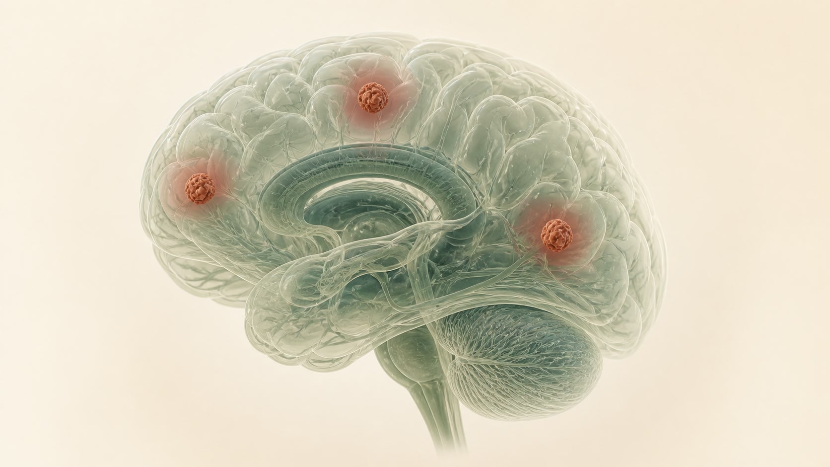
A brain metastasis is a deposit from cancer elsewhere in the body. The number, total volume, location, pressure effect, cancer biology, disease outside the brain, general fitness and personal goals all influence treatment.
01
What brain metastases are
Brain metastases are biologically different from primary brain tumours. Some are found during cancer staging; others present with a seizure, focal deficit or pressure symptoms. The primary cancer and available brain-active systemic treatments matter.
A new brain lesion in a person with cancer is not automatically a metastasis. Infection, treatment effects, vascular lesions and a new primary tumour can sometimes resemble one.
02
Possible symptoms
Symptoms can include a first seizure, new weakness or numbness, language or visual difficulty, impaired balance, cognitive change, worsening headache, vomiting or drowsiness. Small lesions may be asymptomatic; a single lesion with oedema can be very symptomatic.
03
Assessment and tests
- Neurological examination and contrast-enhanced brain MRI when suitable.
- Review of lesion number, total volume, site, oedema and mass effect—not number alone.
- Assessment of disease outside the brain, tumour biology, performance status and available systemic treatment.
- Tissue diagnosis when the origin is uncertain or the result would change treatment.
- NICE NG99 is a UK evidence reference for people aged 16 and over, not a local access or timing guarantee.
04
Non-operative treatment
Stereotactic radiosurgery or radiotherapy can give local control without craniotomy for selected lesions. Whole-brain radiotherapy has a more limited, individual role because effects on cognition and global intracranial control must be balanced. Brain-active systemic treatment may be important for selected cancer subtypes.
Supportive treatment, rehabilitation and symptom control are active parts of care. For some people, avoiding burdensome local treatment is consistent with their goals; this is a shared oncological decision, not abandonment.
05
When surgery may help
Surgery may be useful for a large accessible lesion causing mass effect, when rapid decompression is needed, when tissue diagnosis is important or when the diagnosis is uncertain. It does not treat microscopic or other intracranial disease by itself.
For smaller or deeper lesions, radiosurgery may avoid craniotomy but does not provide immediate mechanical decompression or tissue. No single approach is best for every patient.
06
Your care pathway
- Treat acute pressure or neurological deterioration as an emergency.
- Define brain disease and the wider cancer situation.
- Discuss neurosurgery, radiation oncology and medical oncology together.
- Compare surgery, focused radiation, systemic treatment and supportive care against function and priorities.
- Plan imaging, rehabilitation, seizure support and a route for reassessment if symptoms change.
07
Uncertainty and follow-up
Older trials may not reflect modern radiosurgery or molecular treatments, and average survival figures cannot predict an individual. After radiosurgery or systemic treatment, radiation effect or inflammation can resemble progression.
Follow-up should be coordinated with the wider cancer team and include neurological function, cognition, symptom burden and quality of life as well as imaging.