Research-based patient information · adults; children need separate review
Meningioma: when monitoring or treatment may be appropriate
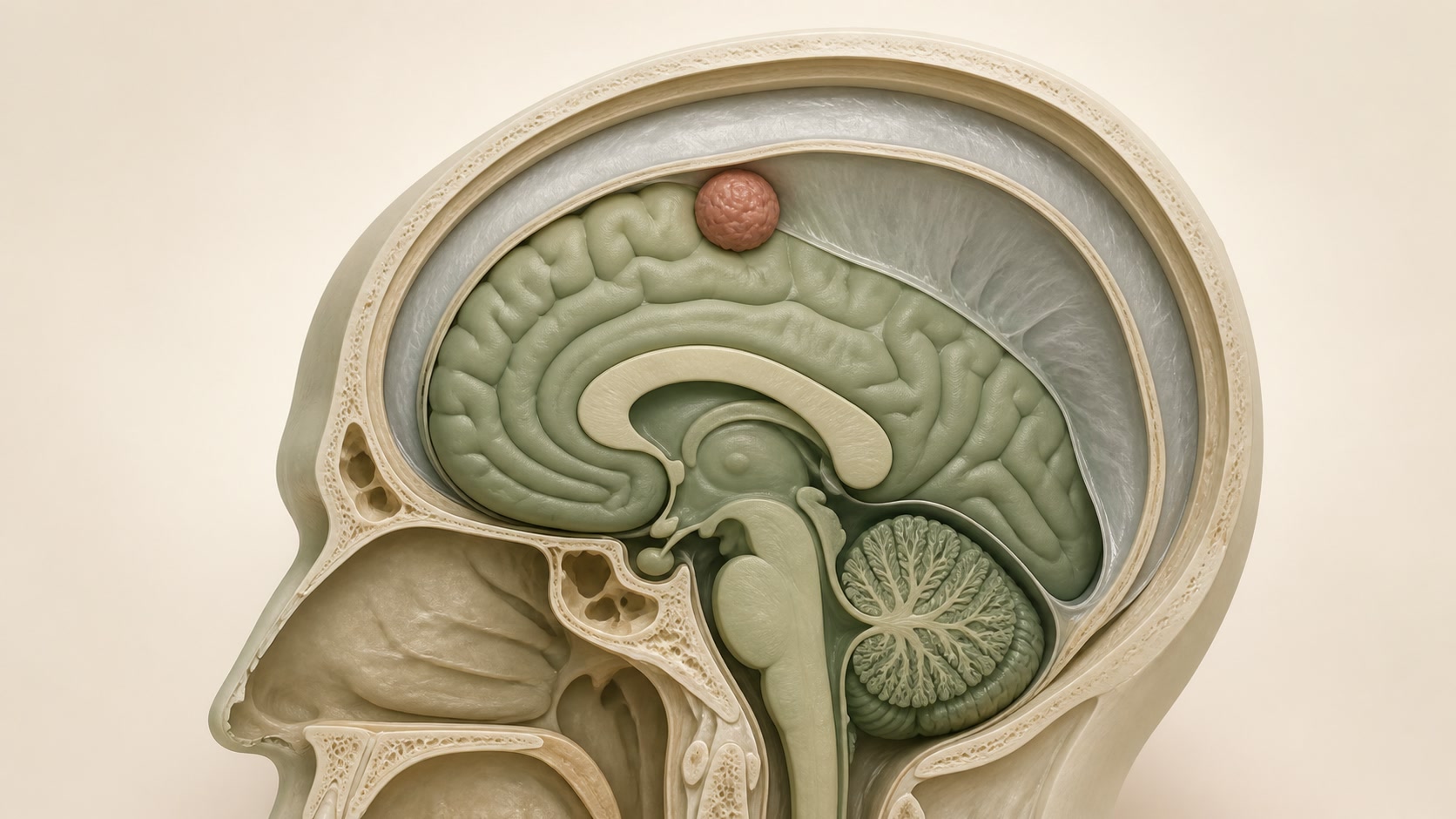
A meningioma usually arises from the coverings of the brain or spinal cord. Many are slow-growing and discovered incidentally. The scan finding, symptoms, growth, location and effect on nearby structures need to be considered together.
01
What a meningioma is
Most meningiomas are not the same as an infiltrating brain cancer, but their behaviour varies. A benign-appearing tumour can still matter if it presses on the brain, spinal cord, optic pathways, cranial nerves or major vessels.
Imaging often suggests the diagnosis, while tissue obtained at surgery provides the definitive grade. Atypical imaging or rapid growth may widen the differential diagnosis.
02
Possible symptoms
Symptoms depend on site and can include seizures, focal weakness or numbness, visual or hearing change, language difficulty, cognitive or personality change, headache from mass effect, or spinal cord and nerve-root symptoms. Many small incidental meningiomas cause no symptoms.
Headache is common in the population. Its presence beside a meningioma does not prove that the tumour is the cause.
03
Assessment and tests
- Neurological and functional examination directed to the tumour's location.
- Contrast-enhanced MRI, with CT where bone change or calcification matters.
- Comparison with earlier imaging to establish growth rather than relying on a single scan.
- Review of oedema, pressure, venous sinus or cranial-nerve involvement and the person's health and priorities.
- NICE NG99 is a UK evidence reference for people aged 16 and over, not a local monitoring guarantee.
04
Active monitoring
Observation is often the first option for an asymptomatic incidental meningioma without threatening features. The interval and duration of imaging are individual and depend on age, size, location, growth pattern, symptoms and whether a future intervention would be feasible.
Radiological growth is not identical to symptom progression. However, documented important growth can change the balance before neurological function is lost.
05
Surgery and radiotherapy
Surgery may be considered for attributable symptoms, clinically important growth, mass effect or oedema, threatened vision or cranial-nerve function, tumour-related seizures, diagnostic uncertainty or features suggesting more aggressive disease. The aim and safe extent depend strongly on attachment to critical structures.
Radiotherapy or stereotactic radiosurgery may be an alternative or complement for selected small, residual, recurrent, inaccessible or higher-grade tumours. Treatment is not automatic for every imaging finding.
06
Your care pathway
- Confirm whether the meningioma is incidental or plausibly explains a symptom.
- Establish location, pressure effects and growth using suitable imaging and comparison scans.
- Discuss monitoring, surgery and focused or fractionated radiotherapy in the appropriate specialist team.
- Balance tumour control against neurological function, operative risk and the person's priorities.
- Agree the next scan, symptom safety-net and rehabilitation or seizure support where needed.
07
Uncertainty and follow-up
Imaging cannot predict the exact growth pattern of one tumour. Surgical series describe selected patients and cannot promise an individual result. After treatment, surveillance depends on grade, extent of removal, site and clinical course.
Useful follow-up asks not only whether the tumour changed, but whether vision, hearing, movement, seizures, cognition and independence changed.