Research-based patient information · adults and children
Head injury: assessment, recovery and the neurosurgical role
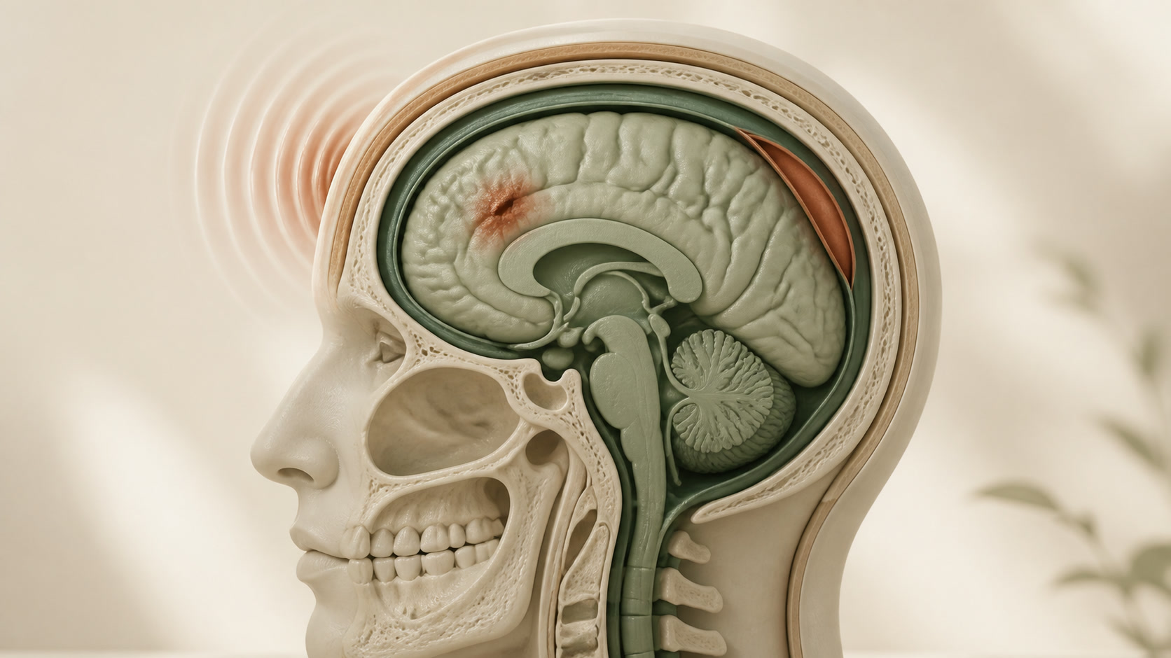
A blow to the head may cause no structural injury, concussion, brain contusion, skull fracture or bleeding around or within the brain. Symptoms can evolve, so assessment uses the event, age, examination and risk factors—not the appearance of the impact alone.
01
What can happen after an impact
The skull and meninges protect the brain, but rapid movement can stretch brain tissue and vessels. A contusion is bruised brain tissue. An epidural, subdural or intracerebral haematoma is a collection of blood in a different anatomical layer or compartment. These injuries can create pressure even when the external wound looks small.
Concussion describes a clinical brain-function disturbance and may occur without an abnormal CT. A normal early scan does not mean every symptom is unreal, while symptoms alone do not identify a haematoma.
02
Adults and older people
Assessment considers loss of consciousness or memory, vomiting, headache, seizure, neurological signs, the injury mechanism, intoxication and previous brain surgery. Age, bleeding disorders and medicines that affect clotting can lower the threshold for imaging or observation.
Some intracranial bleeding, especially subdural bleeding, may present later with worsening headache, confusion, imbalance, weakness or declining function. New deterioration after discharge requires reassessment.
03
Children and people under 16
Children need age-specific observation and CT criteria. In babies and young children, behaviour, feeding, interaction, fontanelle, scalp swelling and the account of what happened are interpreted in developmental context. Concern about possible non-accidental injury requires a safeguarding pathway.
A responsible adult should receive clear written and verbal advice after discharge. Return to school, play and sport should be gradual and guided by symptoms and professional advice; the adult pathway should not be applied automatically.
04
Assessment and tests
- Immediate stabilisation where needed, followed by Glasgow Coma Scale, pupil, cranial-nerve and limb examination.
- CT of the head when age-specific clinical criteria indicate possible important injury; cervical-spine imaging when the mechanism or examination requires it.
- Observation with repeated neurological checks when symptoms, timing or risk make a single assessment insufficient.
- MRI only for selected later questions; it is not usually the first emergency test for clinically important acute bleeding.
- NICE NG232 is a UK evidence reference covering babies, children, young people and adults; it is not a local service-time guarantee.
05
When observation and recovery support may be enough
Many people with a reassuring assessment do not need surgery. Care may include a period of observation, written safety-net advice, clinician-directed symptom treatment, sleep and activity guidance, and a staged return to study, work, driving and sport.
Persistent headache, dizziness, cognitive difficulty, sleep change or emotional symptoms after concussion deserve review and rehabilitation support. Avoiding another head impact during recovery is important; exact restrictions are individual.
06
Persistent symptoms and referral
When problems continue after a head injury, UK NICE NG232 advises considering referral to a clinician trained in assessing and managing the consequences of traumatic brain injury. Depending on the problem, this may be a neurologist, neuropsychologist, clinical psychologist, neurosurgeon or a multidisciplinary neurorehabilitation team.
The referral should match the person's symptoms and age; children need paediatric expertise and may also need coordinated school support. New deterioration is an emergency issue rather than a routine referral.
07
When neurosurgery may be needed
Neurosurgery may evacuate an expanding or pressure-producing haematoma, decompress the brain, control bleeding, repair selected depressed skull fractures or dural leaks, or monitor and manage intracranial pressure. The decision integrates consciousness, neurological change, scan appearance, mass effect, clotting status and the person's overall condition.
A contusion or small bleed is not an automatic operation. Repeat examination or imaging may be safer when the lesion is stable, while rapid clinical or radiological progression can change the decision.
08
A clear patient journey
- Make the scene safe and seek emergency help for red-flag symptoms.
- Record what happened and assess consciousness, neurology, age-specific risk and medicines.
- Choose discharge with safety-netting, observation, CT or specialist escalation according to findings.
- If a significant lesion is present, compare monitoring and supportive care with surgery based on pressure and function.
- Plan recovery, rehabilitation, return to activities and a route back if symptoms persist or worsen.
09
Uncertainty and follow-up
Symptoms and imaging can change after the first assessment, which is why safety-net advice matters. CT is designed to find important acute structural injury, not to measure every effect of concussion or predict an individual recovery time.
Follow-up should address cognition, balance, sleep, mood, headache, work or school and safe return to driving or sport. New deterioration takes priority over a scheduled appointment.