Research-based patient information · children and adults
Hydrocephalus: understanding cerebrospinal-fluid pathways and care
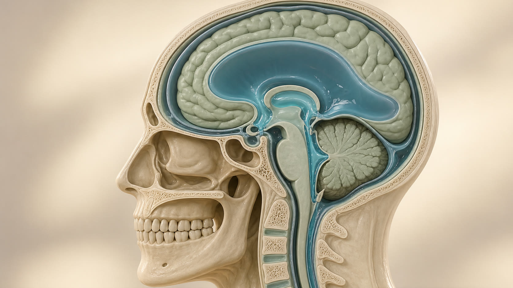
Hydrocephalus is an active disturbance of cerebrospinal-fluid (CSF) circulation that can enlarge the brain's ventricles and affect function. Enlarged ventricles on a scan are a description, not a diagnosis or an automatic reason for surgery.
01
What hydrocephalus means
CSF is produced within and around the brain, flows through connected ventricular spaces and is then absorbed. Hydrocephalus develops when this circulation or absorption is disturbed and the ventricles become pathologically distended. A blockage, previous bleeding, infection, tumour, developmental condition or mixed mechanism may be involved.
Ventricular enlargement can also reflect loss of brain volume, stable congenital anatomy or a compensated state. The diagnosis therefore depends on symptoms, examination, the cause and change over time—not on one measurement alone.
02
Infants and children
In an infant, open skull sutures can allow the head to enlarge. Concerning changes can include head circumference crossing centile lines, a tense fontanelle when calm and upright, downward-looking eyes, repeated vomiting, feeding difficulty, unusual irritability or sleepiness, breathing pauses, or developmental plateau or loss of skills.
Older children may develop progressive or morning headache, vomiting, visual change, abnormal eye movements, deteriorating balance, school performance or alertness. These features have other possible causes, so age-appropriate assessment is essential.
03
Acute hydrocephalus in adults
A sudden blockage or hydrocephalus related to haemorrhage, tumour, infection or a failed CSF-diversion procedure can raise pressure quickly. Declining consciousness or brainstem signs make this a time-critical problem.
Emergency treatment may include temporary ventricular drainage while the cause is assessed. This is different from a planned decision for chronic symptoms.
04
Idiopathic normal-pressure hydrocephalus (iNPH)
iNPH is a chronic adult syndrome in which gait difficulty is usually the most characteristic feature. Short steps, poor foot clearance, hesitation, slow unstable turning and falls may occur. Slowed thinking, reduced attention or initiative and urinary urgency can coexist; the complete traditional triad is not required.
Parkinsonian disorders, dementia, spinal disease, neuropathy, arthritis, vestibular disease, medicines and urological conditions can mimic or accompany iNPH. Ventricular size or a single scan pattern cannot decide whether a shunt will help.
05
Assessment and tests
- History and neurological examination, including alertness, eye movements, vision, gait and balance; serial head measurements and development in infants.
- Ultrasound through an open fontanelle, MRI for CSF pathways and causes, or CT when speed is important in an emergency.
- Comparison with earlier imaging obtained when the person was clinically well; stable ventricular size does not by itself exclude shunt or ETV failure.
- For suspected iNPH: reproducible gait testing, cognitive assessment and, in selected people, a lumbar tap, drainage, infusion study or pressure monitoring. Lumbar puncture is not a test for acute obstructive hydrocephalus.
06
Observation and non-operative care
Stable asymptomatic ventriculomegaly, ventricular enlargement from brain-volume loss and some compensated conditions may be monitored with a defined clinical and imaging plan. Monitoring should include symptoms that would prompt earlier review.
Treating a contributing tumour, infection or haemorrhage may be part of care, but it does not always remove the need for CSF diversion. There is no evidence-based general claim that observation is equivalent to diversion for active, progressive or pressure-threatening hydrocephalus.
07
Shunt and endoscopic treatment
A ventriculoperitoneal shunt can treat obstructive, communicating or mixed hydrocephalus by diverting CSF to the abdomen. Endoscopic third ventriculostomy (ETV) creates an internal bypass and is most suitable for selected obstructive patterns. ETV with choroid-plexus cauterisation may be considered for selected infants in experienced programmes.
Choice depends on age, cause, anatomy, previous infection or bleeding and local expertise. Shunts can obstruct, become infected, disconnect or over- or under-drain; ETV can close, including late. Neither option removes the need for long-term awareness and follow-up.
08
A clear patient journey
- Recognise the clinical pattern and whether it is emergency, urgent or suitable for planned assessment.
- Compare symptoms and examination with age-appropriate imaging and the person's previous baseline.
- Identify the likely cause and discuss observation, cause-directed care, shunt or ETV where relevant.
- Agree realistic aims, material risks, the failure-symptom safety plan and the follow-up team.
- Review development, cognition, walking, independence and quality of life—not ventricular size alone.
09
What remains uncertain
A procedure can control CSF physiology but cannot guarantee reversal of injury caused by the underlying condition. Response—especially in iNPH—and the lifetime risk of further procedures vary between individuals. The most useful discussion separates what is known from the scan, what fits the clinical pattern and what is still uncertain.