Patient information · adults
Lumbar disc herniation: understanding the options
A guide to symptoms, assessment and possible care pathways. It is for adults with a suspected lumbar disc herniation; spinal stenosis, fracture, infection, tumour, deformity and inflammatory disease need separate assessment.
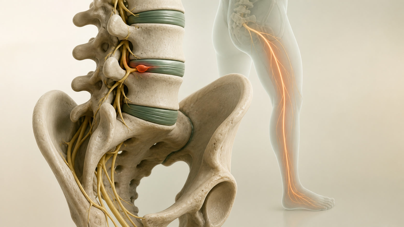
AI-generated educational anatomy and fictional movement footage. It does not depict an actual patient, consultation, diagnosis, procedure or treatment outcome.
01
Pain travelling into a leg is not always nerve-root pain
Back or buttock pain can spread into the leg from muscles, joints, tendons, the hip or sacroiliac region. Nerve-root pain is often sharp, burning, electric or shooting, but symptoms alone cannot prove the cause.
Radiculopathy means there is evidence that a nerve root is not working normally, such as a matching pattern of weakness, altered sensation or reduced reflexes. A musculoskeletal problem can be the main pain source or can coexist with a nerve-root problem.
02
How clinicians decide whether a disc is relevant
A disc finding matters most when four parts agree: the symptoms, the examination, the scan and the clinical course. The side and level of a compressed nerve on MRI should fit the symptoms and examination findings.
An MRI report is not a decision by itself. Disc bulges and degenerative changes are common in people without pain, particularly with increasing age. Conversely, a scan can be useful when it answers a specific clinical question or helps assess a possible emergency.
03
When surgery may be considered
- Emergency assessment and urgent imaging: suspected cauda equina syndrome, including the bladder, bowel, saddle/genital sensation or sexual-function changes described above, or a severe/progressive bilateral neurological deficit. Decompression is considered only when the clinical syndrome and compressive imaging findings correspond.
- Urgent: new or worsening objective weakness, including functionally important foot drop, needs urgent same-day clinical assessment. The timing of MRI and specialist review depends on the findings.
- Elective: persistent, disabling—usually leg-dominant—nerve-root symptoms despite a reasonable period of non-operative care, when symptoms, examination and MRI are concordant.
- Stable tingling or numbness on its own is not automatically an emergency, but it should be reassessed if it progresses, comes with weakness, or is accompanied by bladder, bowel or saddle symptoms.
04
What non-operative care can involve
Without an emergency syndrome or progressive major weakness, many people improve with time. Care may include explanation, maintaining activity as tolerated, tailored pain management, rehabilitation and attention to other possible contributors such as hip, tendon, joint or muscular problems.
Some disc herniations reduce in size on later imaging, especially when material has moved beyond the disc. That does not predict an individual person’s recovery and repeat imaging does not reliably measure how well someone feels.
05
What discectomy can and cannot do
Discectomy or microdiscectomy removes disc material that is compressing a matching nerve root. Its most predictable aim is relief of concordant radiating leg pain or protection of an endangered nerve function—not treatment of every kind of back pain or every age-related MRI change.
Back pain may improve, remain unchanged or sometimes persist. Risks can include dural tear and spinal-fluid leak, infection, bleeding, nerve injury, ongoing neuropathic symptoms, recurrent disc herniation and a need for further treatment. The relevant risks depend on the person and the operation being discussed.
06
Surgery compared with a conservative-first pathway
Across trials, surgery generally brings faster relief of leg pain and a faster sense of recovery for appropriately selected people. By one to several years, average differences in pain and disability often become much smaller.
This does not mean that non-operative care is generally better in the long term, or that surgery is unnecessary. Many studies allowed people initially treated without surgery to have delayed surgery if needed. The choice is about urgency, symptoms, function, examination, imaging, trajectory and the person’s preferences.
07
Questions worth taking to a consultation
- Do my symptoms and examination findings match the level and side shown on the scan?
- What else could be contributing to the pain or weakness?
- Is there a reason this needs emergency, urgent or planned assessment?
- What improvement is realistic for leg pain, numbness, weakness and back pain separately?
- What are the alternatives, the material risks and the follow-up plan in my situation?